
Monitoring and managing kidney swelling detected before birth — a condition that often resolves on its own, but requires careful post-natal follow-up to protect kidney health.
Antenatal hydronephrosis (ANH) is a dilation of the renal pelvis — the collecting area of the kidney — detected on a routine antenatal (pregnancy) ultrasound. It is the most common abnormality identified on prenatal scanning, occurring in around 1–5% of pregnancies.
The cause ranges from a transient, physiological dilation that resolves after delivery to a structural blockage or vesicoureteral reflux (VUR) requiring treatment. A post-natal evaluation is essential in all cases to determine which babies need monitoring, prophylactic antibiotics, or referral for surgical assessment — and which can safely be watched with periodic ultrasound scans alone.
The Society for Fetal Urology (SFU) grading system (1–4) helps classify severity and decide how urgently post-natal investigation is needed.
The majority of antenatally detected hydronephrosis cases resolve on their own — a calm, structured follow-up plan is usually all that is needed.
The first post-natal ultrasound is usually done at 4–7 days of age (not at birth, when neonatal dehydration can underestimate dilation).
Most babies with ANH are clinically well. These warning signs require immediate attention.
Note: If your baby was diagnosed with hydronephrosis before birth, ensure a post-natal nephrology review is scheduled — even if the baby seems perfectly healthy at delivery.
Any fever in a baby under 3 months with a known history of antenatal hydronephrosis must be evaluated urgently for a UTI or obstructive infection.
A significant bilateral obstruction can impair kidney drainage. Reduced wet nappies in the first days of life need investigation.
A very dilated kidney can occasionally be felt as a lump in the flank — always requires urgent imaging.
A large pelvic diameter (APD >15mm in the third trimester) or calyceal dilation visible before birth warrants early post-natal investigation.
Tailored to the grade, cause and progression of the hydronephrosis.
A kidney ultrasound at 4–7 days of life to confirm the diagnosis, measure pelvic dilation and classify the grade of hydronephrosis.
MCUG (to detect VUR), MAG3 renogram (to assess drainage and differential function) or DMSA scan ordered based on grade and clinical picture.
Mild cases: serial ultrasound until resolution. Moderate: antibiotic prophylaxis and regular monitoring. Severe or obstructive: referral to paediatric urology for surgical intervention.
The majority of antenatally detected cases (especially low-grade ones) resolve on their own after birth. The finding prompts a structured follow-up plan — it does not mean your baby will need surgery or has serious kidney disease. A calm, scheduled post-natal review is the right first step.
Surgery is only needed if the hydronephrosis worsens over time, if kidney function is compromised on a MAG3 scan, or if there are repeated infections. Most decisions are made after the 4–6 week post-natal ultrasound and any additional imaging required.
Low-dose prophylactic antibiotics are commonly prescribed for moderate to high-grade ANH, or when VUR is suspected, to reduce the risk of a UTI while investigations are ongoing. This decision is individualised based on the grade and imaging findings.
Mild cases that resolve on the first ultrasound may require just one or two additional scans. Moderate or persistent cases are followed for 12–24 months or until resolution is confirmed. High-grade or structural cases may need longer-term nephrology and urology follow-up.
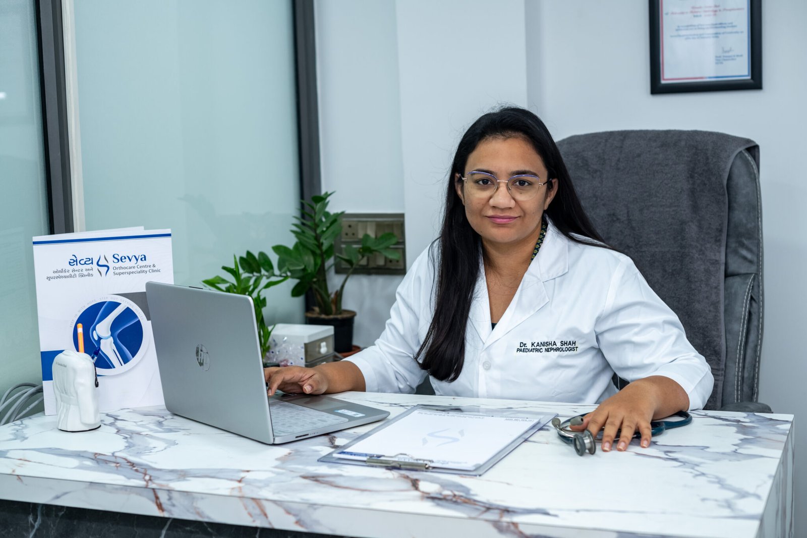
Nurturing Young Kidneys for a Lifetime
When it comes to your child's kidney health, finding the right specialist makes all the difference. As the only dedicated child kidney doctor in East Ahmedabad and one of the select few across Gujarat, Dr. Kanisha Shah brings rare, highly specialized expertise directly to your community. She provides precise, deeply reassuring care for complex conditions—from persistent bed-wetting and recurrent UTIs to nephrotic syndrome. Dr. Shah partners closely with parents, delivering honest, evidence-based treatments designed to protect, heal, and nurture developing kidneys for a lifetime.
Have a question about your child's condition, or want to schedule a visit?